How Does Gluten Activate T Cells?

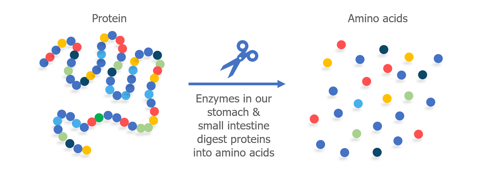
All proteins are made up of smaller parts called amino acids. The amino acids are linked together to form a long chain, like beads that have been strung together to form a necklace. 

When we eat protein, enzymes in our digestive system (called proteases) break apart the chain into individual amino acids. In our intestine, cells called enterocytes absorb the amino acids so they can be used to fuel our bodies and rebuild our own proteins.
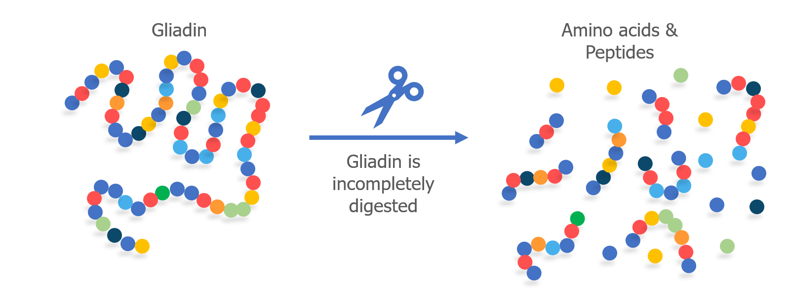
When humans eat gluten, it is not completely digested into individual amino acids in the small intestine. Gliadin, specifically, is difficult for our bodies to digest because it contains a high proportion of two amino acids—proline and glutamine. Because it is only partially digested, small fragments of the gliadin protein chain remain as peptides. This happens in people with and without celiac disease.
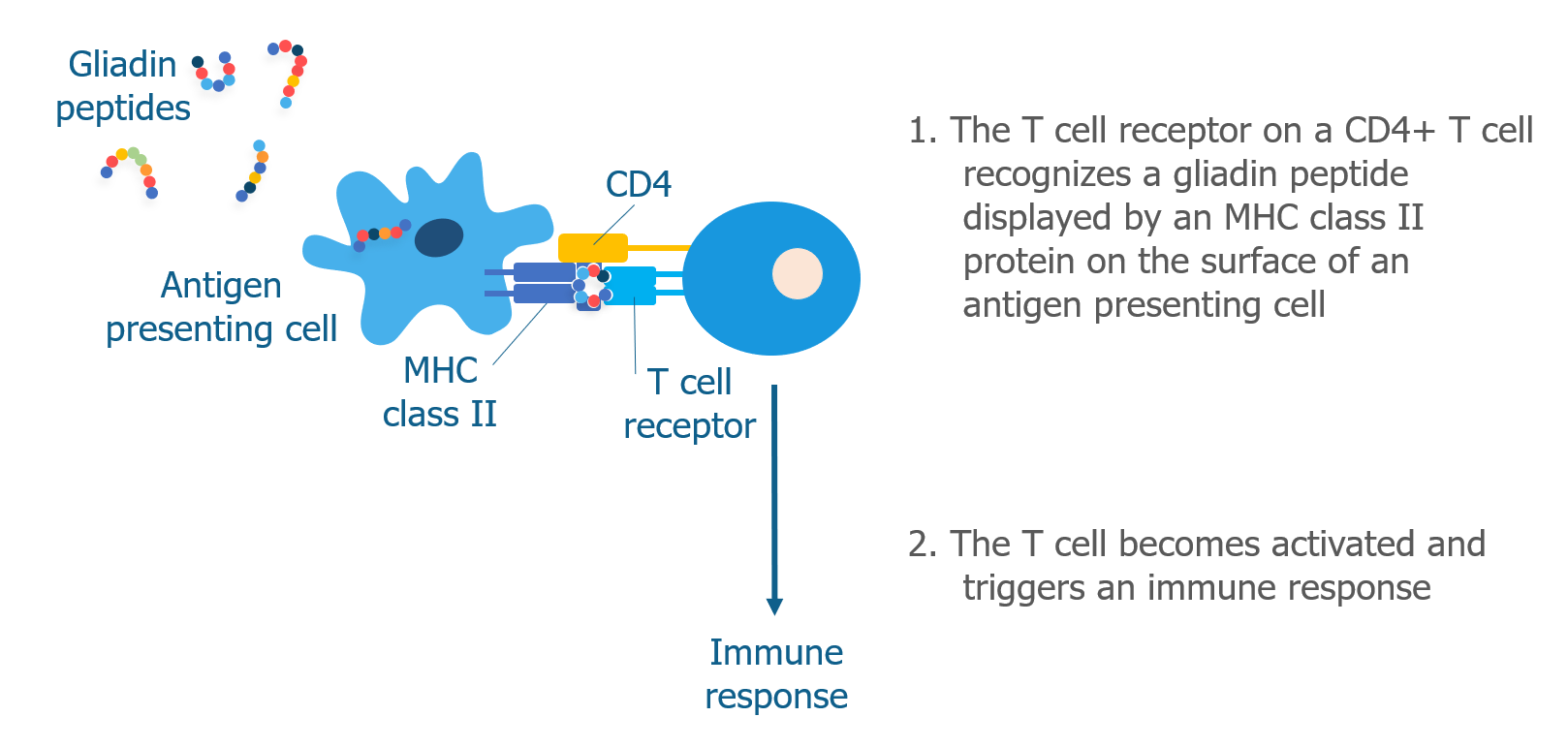
Antigen presenting cells located in the intestine take up the undigested gliadin peptides and display them on their surface with an MHC class II molecule. CD4+ T cells with receptors shaped like “locks” that match gliadin peptide “keys” mistakenly recognize gluten as a harmful pathogen and become activated. This begins the immune response.
Once CD4+ T cells become activated, they can produce different subsets of helper T cells (as described here). In celiac disease, the most common type of helper T cell produced is Th1, which produces cytokines, such as interferon gamma, that recruit other immune cells and cause tissue damage. There is also evidence for Th17 cells in celiac disease. Th17 cells produce interleukin-17, which is a key cytokine involved in several different autoimmune diseases.1

<< PREVIOUS                                                                                                                                         NEXT >>

REFERENCES
  1. Jabri B, Sollid LM. T Cells in Celiac Disease. J Immunol. 2017;198(8):3005-3014.
GLOSSARY

Amino acids – Small units that are linked together by bonds to form proteins.

Antigen presenting cell – A specialized immune cell that presents peptides to CD4+ or CD8+ T cells. Peptides are presented by MHC I or MHC II proteins.

Cytokines – Small proteins that are made and released by immune cells. Allows cells to send signals and provide instructions to other cells.

Enterocytes – Cells that make up the intestinal lining. Responsible for absorbing nutrients.

Gliadin – One of the wheat proteins that forms gluten. Responsible for triggering an immune response in patients with celiac disease.

Gluten – A type of protein found in cereal grains. Wheat gluten is made of two proteins called gliadin and glutenin.

Helper T cell – Adaptive CD4+ immune cell that produces cytokines when activated.

MHC class II protein – Major histocompatibility complex class II protein. Found on the surface of antigen presenting cells. Presents peptides to CD4+ T cells.

Peptide – A small protein fragment consisting of a chain of amino acids.

Protease – A type of enzyme that breaks up proteins into smaller parts.

Receptor – A protein that is located on the surface of a cell and interacts with other proteins. Receptors act as the “locks” that recognize specific pathogen “keys”.

T cell – A type of adaptive immune cell. Also called T lymphocyte.


RELATED

Share this Post

What Are the Different Types of T Cells?

There are 3 main types of T cells: cytotoxic, helper, and regulatory. Each of them has a different role in the immune response.

Cytotoxic T cells (CD8+)

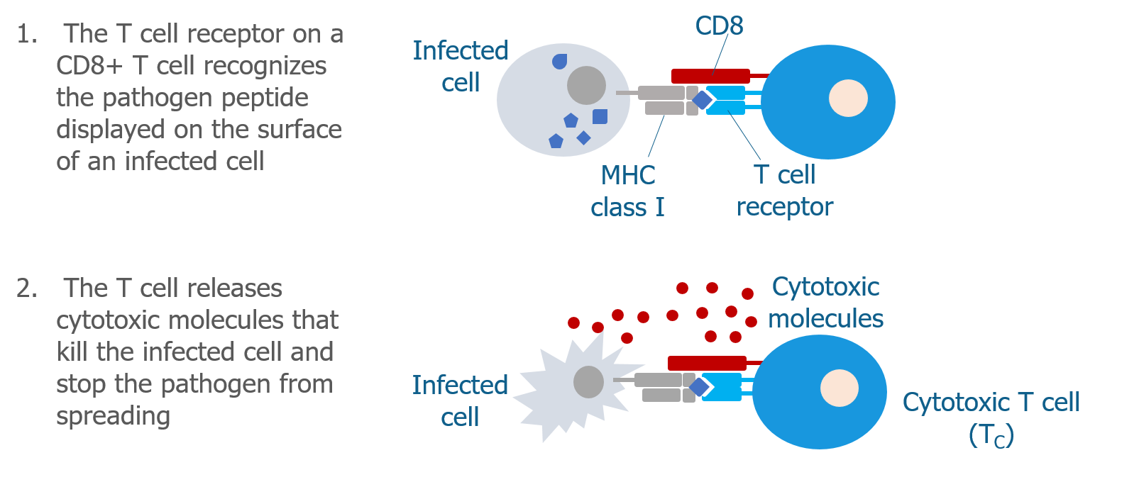
Cytotoxic T cells (Tc cells) have a co-receptor called CD8 on their cell surface. CD8 partners with the T cell receptor and with MHC class I molecules, acting as a sort of bridge. This bridge allows cytotoxic T cells to recognize normal cells that are infected by a pathogen. When the cytotoxic T cell recognizes the infected cell, it becomes activated and produces molecules that kill the infected cell, destroying the pathogen in the process.

Helper T cells (CD4+)

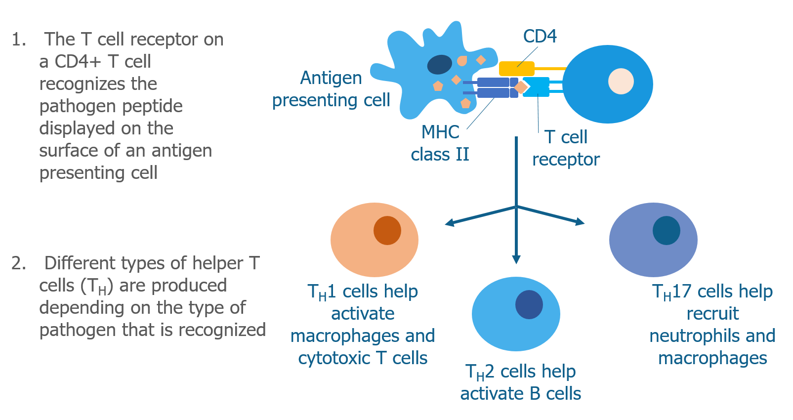
Helper T cells (Th cells) have a different co-receptor called CD4 on their cell surface. CD4 also partners with the T cell receptor but interacts with MHC class II molecules instead of MHC class I molecules. This allows helper T cells to recognize pathogen peptides that have been displayed by antigen presenting cells. When helper T cells recognize a peptide on an antigen presenting cell, they become activated and begin to produce molecules called cytokines that signal to other immune cells.

There are many subtypes of helper T cells (ie, Th1, Th2, Th17). Each subtype produces a specialized combination of cytokines that depends on type of pathogen that the helper T cell has recognized—some cytokines are more effective than others in the process of eliminating certain invaders.

Regulatory T cells

Regulatory T cells (Treg cells) also have CD4 on their surface, but they do not activate the immune system like helper T cells do. Instead, regulatory T cells play a protective role by shutting off the immune response when it is no longer needed. This prevents excessive damage to the normal cells and tissues in the body. Regulatory T cells suppress the immune response in several ways, including:

  • Producing anti-inflammatory cytokines that suppress the immune response
  • Releasing molecules that kill activated immune cells
  • Changing the way dendritic cells behave so they can’t activate T cells

Which T cells are involved in celiac disease?

Of the 3 types of T cells described above, the CD4+ T helper cells play a leading role in celiac disease. These are the cells that mistakenly recognize gluten as a pathogen and trigger an immune response (see below).1 However, cytotoxic CD8+ T cells known as intraepithelial lymphocytes (IELs) are also important as they are drivers of tissue damage in the intestine.2

<< PREVIOUS                                                                                                                                         NEXT >>

REFERENCES
  1. Jabri B, Sollid LM. T Cells in Celiac Disease. J Immunol. 2017;198(8):3005-3014.
  2. Abadie V, Discepolo V, Jabri B. Intraepithelial lymphocytes in celiac disease immunopathology. Semin Immunopathol. 2012;34(4):551-566.
GLOSSARY

Antibodies – Y-shaped proteins that recognize foreign pathogens. Made by B cells. Also called immunoglobulins.

Antigen presenting cell – A specialized immune cell that presents peptides to CD4+ or CD8+ T cells. Peptides are presented by MHC I or MHC II proteins.

CD4 – A co-receptor on the surface of helper T cells.

CD8 – A co-receptor on the surface of cytotoxic T cells.

Cytokines – Small proteins that are made and released by immune cells. Allows cells to send signals and provide instructions to other cells.

Cytotoxic T cell – Adaptive CD8+ immune cell that kill infected cells when activated.

Dendritic cell – A type of antigen presenting cell that processes pathogens and foreign proteins. Presents peptides to T cells.

Helper T cell – Adaptive CD4+ immune cell that produces cytokines when activated.

Immune cells – Specialized white blood cells (also called leukocytes) that fight infection.

Intraepithelial lymphocytes – Cytotoxic CD8+ T cells that reside in the intestine. Contribute to tissue damage in celiac disease.

MHC class I protein – Major histocompatibility complex class I protein. Found on the surface of normal cells. Presents peptides to CD8+ T cells.

MHC class II protein – Major histocompatibility complex class II protein. Found on the surface of antigen presenting cells. Presents peptides to CD4+ T cells.

Pathogens – Bacteria and viruses that can cause disease.

Peptide – A small protein fragment consisting of a chain of amino acids.

Receptor – A protein that is located on the surface of a cell and interacts with other proteins. Receptors act as the “locks” that recognize specific pathogen “keys”.

Regulatory T cell – An adaptive immune cell that suppresses the immune response.

T cell – A type of adaptive immune cell. Also called T lymphocyte.


RELATED

Share this Post

T Cells

Like all immune cells, T cells have receptors (in this case called T cell receptors) that recognize antigens. Once the antigen is recognized, the T cell becomes activated. Activated T cells are responsible for destroying the pathogen, either directly by killing the infected cell, or indirectly by activating B cells or innate immune cells—this is often called the cell-mediated immune response.

How do T cells recognize antigens?

Unlike other immune cells, T cells are not able to recognize antigens on their own. The antigens have to be presented to them in a way that allows the T cell receptor to recognize them.

Antigens can be presented to T cells in one of two ways: 1) by normal cells that are infected; or 2) by professional antigen presenting cells.

Infected cells

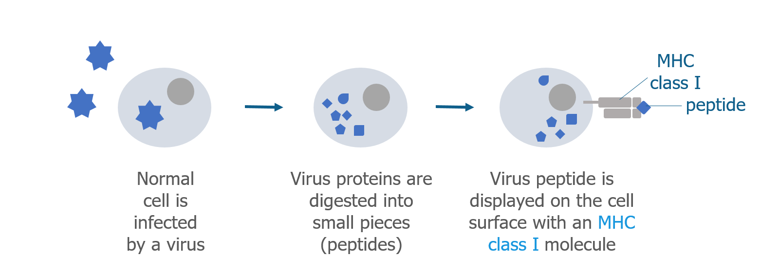
Cells normally break down proteins and make new ones to replace them. Many cellular proteins are broken down into smaller pieces called peptides. Some of these peptides are sent to the surface of the cell along with a major histocompatibility complex (MHC) class I protein. The MHC class I protein displays the peptide on the outside of the cell so it can be recognized by certain T cells. If a cell has been infected by a foreign pathogen, such as a virus that lives inside the cell, viral peptides (antigens) will be displayed on the surface of the infected cell.

Antigen presenting cells

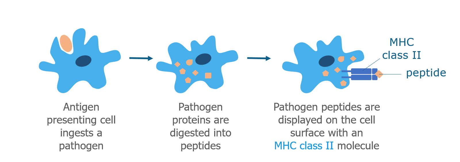
Dendritic cells, macrophages, and B cells are professional antigen presenting cells that process pathogens. When an antigen presenting cell bumps into a pathogen, it brings it inside the cell using a process called phagocytosis (cell eating). Similar to normal cellular proteins, the pathogen is digested into peptides, which are displayed on the surface of the cell together with a MHC class II protein. Like the MHC class I protein, the MHC class II protein displays the peptide (antigen) and interacts with certain T cells.

<< PREVIOUS                                                                                                                                         NEXT >>

GLOSSARY

Antigen – A protein that is recognized by an immune cell receptor and triggers an immune response.

Antigen presenting cell – A specialized immune cell that presents peptides to CD4+ or CD8+ T cells. Peptides are presented by MHC I or MHC II proteins.

B cell – A type of adaptive immune cell. Also called B lymphocyte.

Cell-mediated immune response – Immune response that involves the direct or indirect activation of immune cells.

Immune cells – Specialized white blood cells (also called leukocytes) that fight infection.

Innate immune cells – Nonspecific immune cells. Recognize many pathogens. First responders to infection.

MHC class I protein – Major histocompatibility complex class I protein. Found on the surface of normal cells. Presents peptides to CD8+ T cells.

MHC class II protein – Major histocompatibility complex class II protein. Found on the surface of antigen presenting cells. Presents peptides to CD4+ T cells.

Pathogens – Bacteria and viruses that can cause disease.

Peptide – A small protein fragment consisting of a chain of amino acids.

Receptor – A protein that is located on the surface of a cell and interacts with other proteins. Receptors act as the “locks” that recognize specific pathogen “keys”.

T cell – A type of adaptive immune cell. Also called T lymphocyte.


RELATED

Share this Post

The Immune System in Celiac Disease

In the 1960s and 1970s, researchers began to understand that the immune system was involved in celiac disease, and that the immune response occurred upon exposure to gluten.1

One of the first clues was that more lymphocytes were found in the intestines of patients with untreated celiac disease and fewer lymphocytes were found in the intestines of patients who were following a strict gluten-free diet. Another clue to the importance of the immune system in celiac disease was the discovery that patients with celiac disease had antibodies to wheat proteins circulating in their bloodstream.2

By the 1980s and 1990s, researchers understood that T cells (adaptive immune cells) were involved in the immune response to gluten and the development of celiac disease.3-5 They also understood that B cells made antibodies that recognized a normal protein in the intestine called tissue transglutaminase or tTG.6

In the next sections we will learn more about the roles that T cells and B cells play in celiac disease.

T Cells

Like all immune cells, T cells have receptors (in this case called T cell receptors) that recognize antigens. Once the antigen is recognized, the T cell becomes activated. Activated T cells are responsible for destroying the pathogen, either directly by killing the infected cell, or indirectly by activating B cells or innate immune cells—this is often called the cell-mediated immune response.

How do T cells recognize antigens?

Unlike other immune cells, T cells are not able to recognize antigens on their own. The antigens have to be presented to them in a way that allows the T cell receptor to recognize them.

Antigens can be presented to T cells in one of two ways: 1) by normal cells that are infected; or 2) by professional antigen presenting cells.

Infected cells

Cells normally break down proteins and make new ones to replace them. Many cellular proteins are broken down into smaller pieces called peptides. Some of these peptides are sent to the surface of the cell along with a major histocompatibility complex (MHC) class I protein. The MHC class I protein displays the peptide on the outside of the cell so it can be recognized by certain T cells. If a cell has been infected by a foreign pathogen, such as a virus that lives inside the cell, viral peptides (antigens) will be displayed on the surface of the infected cell.

Antigen presenting cells

Dendritic cells, macrophages, and B cells are professional antigen presenting cells that process pathogens. When an antigen presenting cell bumps into a pathogen, it brings it inside the cell using a process called phagocytosis (cell eating). Similar to normal cellular proteins, the pathogen is digested into peptides, which are displayed on the surface of the cell together with a MHC class II protein. Like the MHC class I protein, the MHC class II protein displays the peptide (antigen) and interacts with certain T cells.

What Are the Different Types of T Cells?
Cytotoxic T cells (CD8+)

Cytotoxic T cells (Tc cells) have a co-receptor called CD8 on their cell surface. CD8 partners with the T cell receptor and with MHC class I molecules, acting as a sort of bridge. This bridge allows cytotoxic T cells to recognize normal cells that are infected by a pathogen. When the cytotoxic T cell recognizes the infected cell, it becomes activated and produces molecules that kill the infected cell, destroying the pathogen in the process.

Helper T cells (CD4+)

Helper T cells (Th cells) have a different co-receptor called CD4 on their cell surface. CD4 also partners with the T cell receptor but interacts with MHC class II molecules instead of MHC class I molecules. This allows helper T cells to recognize pathogen peptides that have been displayed by antigen presenting cells. When helper T cells recognize a peptide on an antigen presenting cell, they become activated and begin to produce molecules called cytokines that signal to other immune cells.

There are many subtypes of helper T cells (ie, Th1, Th2, Th17). Each subtype produces a specialized combination of cytokines that depends on type of pathogen that the helper T cell has recognized—some cytokines are more effective than others in the process of eliminating certain invaders.

Regulatory T cells

Regulatory T cells (Treg cells) also have CD4 on their surface, but they do not activate the immune system like helper T cells do. Instead, regulatory T cells play a protective role by shutting off the immune response when it is no longer needed. This prevents excessive damage to the normal cells and tissues in the body. Regulatory T cells suppress the immune response in several ways, including:

  • Producing anti-inflammatory cytokines that suppress the immune response
  • Releasing molecules that kill activated immune cells
  • Changing the way dendritic cells behave so they can’t activate T cells
Which T cells are involved in celiac disease?

Of the 3 types of T cells described above, the CD4+ T helper cells play a leading role in celiac disease. These are the cells that mistakenly recognize gluten as a pathogen and trigger an immune response (see below).7 However, cytotoxic CD8+ T cells known as intraepithelial lymphocytes (IELs) are also important as they are drivers of tissue damage in the intestine.8

How Does Gluten Activate T Cells?

All proteins are made up of smaller parts called amino acids. The amino acids are linked together to form a long chain, like beads that have been strung together to form a necklace. 

When we eat protein, enzymes in our digestive system (called proteases) break apart the chain into individual amino acids. In our intestine, cells called enterocytes absorb the amino acids so they can be used to fuel our bodies and rebuild our own proteins.

When humans eat gluten, it is not completely digested into individual amino acids in the small intestine. Gliadin, specifically, is difficult for our bodies to digest because it contains a high proportion of two amino acids—proline and glutamine. Because it is only partially digested, small fragments of the gliadin protein chain remain as peptides. This happens in people with and without celiac disease.

Antigen presenting cells located in the intestine take up the undigested gliadin peptides and display them on their surface with an MHC class II molecule. CD4+ T cells with receptors shaped like “locks” that match gliadin peptide “keys” mistakenly recognize gluten as a harmful pathogen and become activated. This begins the immune response.

Once CD4+ T cells become activated, they can produce different subsets of helper T cells (as described here). In celiac disease, the most common type of helper T cell produced is Th1, which produces cytokines, such as interferon gamma, that recruit other immune cells and cause tissue damage. There is also evidence for Th17 cells in celiac disease. Th17 cells produce interleukin-17, which is a key cytokine involved in several different autoimmune diseases.7

NEXT TOPIC: What does it mean to be genetically predisposed?

BACK TO MAIN PAGE

REFERENCES
  1. Ferguson A, Murray D. Quantitation of intraepithelial lymphocytes in human jejunum. Gut. 1971;12(12):988-994.
  2. Kowlessar OD. Effect of wheat proteins in celiac disease. Gastroenterology. 1967;52(5):893-897.
  3. Halstensen TS, Scott H, Farstad IN, Michaelsen TE, Brandtzaeg P. In situ two- and three-color immunofluorescence staining of mucosal T-cells in celiac disease. Increase of TCR gamma/delta+CD8- and TCR alpha/beta+CD45R0+ intraepithelial lymphocytes and IL-2R+ TCR alpha/beta+CD4+CD45R0+ lamina propria lymphocytes. Prog Histochem Cytochem. 1992;26(1-4):201-210.
  4. Marsh MN. Gluten, major histocompatibility complex, and the small intestine. A molecular and immunobiologic approach to the spectrum of gluten sensitivity (‘celiac sprue’). Gastroenterology. 1992;102(1):330-354.
  5. Selby WS, Janossy G, Bofill M, Jewell DP. Lymphocyte subpopulations in the human small intestine. The findings in normal mucosa and in the mucosa of patients with adult coeliac disease. Clin Exp Immunol. 1983;52(1):219-228.
  6. Dieterich W, Ehnis T, Bauer M, et al. Identification of tissue transglutaminase as the autoantigen of celiac disease. Nat Med. 1997;3(7):797-801.
  7. Jabri B, Sollid LM. T Cells in Celiac Disease. J Immunol. 2017;198(8):3005-3014.
  8. Abadie V, Discepolo V, Jabri B. Intraepithelial lymphocytes in celiac disease immunopathology. Semin Immunopathol. 2012;34(4):551-566.
GLOSSARY

Adaptive immune cells – Immune cells that recognize specific pathogens. Allow the immune system to “remember” pathogens in case of future infection.

Amino acids – Small units that are linked together by bonds to form proteins.

Antibodies – Y-shaped proteins that recognize foreign pathogens. Made by B cells. Also called immunoglobulins.

Antigen – A protein that is recognized by an immune cell receptor and triggers an immune response.

Antigen presenting cell – A specialized immune cell that presents peptides to CD4+ or CD8+ T cells. Peptides are presented by MHC I or MHC II proteins.

B cell – A type of adaptive immune cell. Also called B lymphocyte.

CD4 – A co-receptor on the surface of helper T cells.

CD8 – A co-receptor on the surface of cytotoxic T cells.

Cell-mediated immune response – Immune response that involves the direct or indirect activation of immune cells.

Cytokines – Small proteins that are made and released by immune cells. Allows cells to send signals and provide instructions to other cells.

Cytotoxic T cell – Adaptive CD8+ immune cell that kill infected cells when activated.

Enterocytes – Cells that make up the intestinal lining. Responsible for absorbing nutrients.

Gliadin – One of the wheat proteins that forms gluten. Responsible for triggering an immune response in patients with celiac disease.

Gluten – A type of protein found in cereal grains. Wheat gluten is made of two proteins called gliadin and glutenin.

Helper T cell – Adaptive CD4+ immune cell that produces cytokines when activated.

Immune cells – Specialized white blood cells (also called leukocytes) that fight infection.

Innate immune cells – Nonspecific immune cells. Recognize many pathogens. First responders to infection.

Intraepithelial lymphocytes – Cytotoxic CD8+ T cells that reside in the intestine. Contribute to tissue damage in celiac disease.

Lymphocyte – Adaptive immune cells. Can by B or T lymphocytes.

MHC class I protein – Major histocompatibility complex class I protein. Found on the surface of normal cells. Presents peptides to CD8+ T cells.

MHC class II protein – Major histocompatibility complex class II protein. Found on the surface of antigen presenting cells. Presents peptides to CD4+ T cells.

Pathogens – Bacteria and viruses that can cause disease.

Peptide – A small protein fragment consisting of a chain of amino acids.

Protease – A type of enzyme that breaks up proteins into smaller parts.

Receptor – A protein that is located on the surface of a cell and interacts with other proteins. Receptors act as the “locks” that recognize specific pathogen “keys”.

Regulatory T cell – An adaptive immune cell that suppresses the immune response.

T cell – A type of adaptive immune cell. Also called T lymphocyte.

Tissue transglutaminase (tTG) – An enzyme that deamidates gliadin peptides causing them to be more immunogenic. Also the target of auto-antibodies in patients with celiac disease.

Share this Post

What Is Gluten?

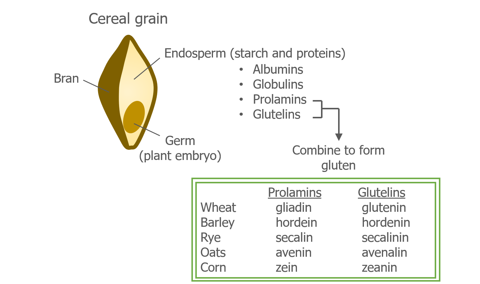
A cereal grain (wheat, barley, rye, oats, corn) is essentially a seed that can germinate and grow into a plant. Each grain has distinct parts: the bran is the outer protective layer, the germ is the plant embryo, and the endosperm contains starch and proteins that provide the nutrients that the germ needs to grow.

There are 4 main types of proteins in the endosperm—albumins, globulins, prolamins, and glutelins. Gluten is made up of 2 of these proteins: prolamins and glutelins. In wheat, the prolamin is called gliadin and the glutelin is called glutenin. Barley, rye, oats, and corn also have prolamins and glutelins, but they have different names.

Although prolamins and glutelins are also found in oats and corn, they are different from those in wheat, barley, and rye and are safe for people with celiac disease. Oats, however, often come into contact with wheat because they are grown in the same fields or harvested with shared equipment. For this reason, it is important to check that the oats you eat are gluten free, which means they were grown and harvested according to a purity protocol.

Gluten is formed when water is mixed with flour. Adding water to dry wheat flour causes gliadin and glutenin to become flexible and begin to stick to each other. When this happens, they form gluten; a very stretchy and elastic substance. The stretchiness and elasticity of gluten, and its ability to trap small pockets of air, make it is useful for baking cakes, breads, and pizza dough.

The link between celiac disease and wheat was discovered by Willem Dicke, a Dutch pediatrician, and was first reported in the 1940’s and 1950’s.1,2 Dr. Dicke noted that the symptoms of celiac disease improved when wheat flour was removed from the diet and got worse when it was reintroduced.

Dr. Dicke and his team of researchers later found that it was wheat protein (gluten), and not wheat starch, that was responsible for the symptoms of celiac disease.2 When they tested different types of wheat proteins individually, they found that gliadin played a main role in triggering the symptoms.3

NEXT TOPIC: The immune system in celiac disease

BACK TO MAIN PAGE

REFERENCES
  1. Dicke W. Simple dietary treatment for the syndrome of Gee-Herter. Ned Tijdschr Geneeskd. 1941;85:17154.
  2. Dicke WK, Weijers HA, Van De Kamer JH. Coeliac disease. II. The presence in wheat of a factor having a deleterious effect in cases of coeliac disease. Acta Paediatr. 1953;42(1):34-42.
  3. Van De Kamer JH, Weijers HA, Dicke WK. Coeliac disease. IV. An investigation into the injurious constituents of wheat in connection with their action on patients with coeliac disease. Acta Paediatr. 1953;42(3):223-231.
GLOSSARY

Gliadin  – One of the wheat proteins that forms gluten. Responsible for triggering an immune response in patients with celiac disease.

Gluten – A type of protein found in cereal grains. Wheat gluten is made of two proteins called gliadin and glutenin.

Glutenin – One of the wheat proteins that forms gluten.

Purity Protocol – A method of growing and harvesting oats that farmers follow to prevent cross-contact with gluten-containing grains (wheat, barley, rye).

Share this Post

What Is Autoimmune Disease?

The immune system is incredibly powerful and capable of causing damage to our own cells while attempting to get rid of pathogens. To make sure this doesn’t happen, many safeguards are in place to guide the immune cells toward pathogens that may cause harm, and away from the thousands of normal proteins that are found in our bodies and in our food.

If these safeguards malfunction, the immune system may recognize a normal, unharmful protein as a threat. In some situations, this can lead to autoimmune disease.

The prefix auto– means “oneself”. Autoimmune disease occurs when the immune system mistakenly recognizes a normal protein in the body as an invader and creates an immune response. Repeated attacks cause inflammation and damage to the tissue where that protein is normally found. As damage accumulates over time, the tissue becomes unable to do its job.

For example, in type I diabetes, the immune system specifically attacks and destroys insulin-producing cells in the pancreas. When these cells are damaged or lost they can no longer make the amount of insulin the body needs.

Over 80 different autoimmune diseases have been identified.1  In most cases, the cause of the autoimmune disease is not known. Researchers think many different factors—such as our genes, environment, illnesses, and infections—play a part in triggering the disease process. 

Celiac disease is an autoimmune disease that affects approximately 1% of the population.2  In contrast to many other autoimmune diseases, celiac disease has a known trigger—a protein called gluten, which is found in cereal grains such as wheat.

NEXT TOPIC: What is gluten?

BACK TO MAIN PAGE

References
  1. Hayter SM, Cook MC. Updated assessment of the prevalence, spectrum and case definition of autoimmune disease. Autoimmun Rev. 2012;11(10):754-765.
  2. Fasano A, Catassi C. Clinical practice. Celiac disease. N Engl J Med. 2012;367(25):2419-2426.
GLOSSARY

Autoimmune disease – May occur when the immune system mistakenly recognizes a normal protein in the body as an invader and creates an immune response.

Gluten – A type of protein found in cereal grains. Wheat gluten is made of two proteins called gliadin and glutenin.

Immune cells – Specialized white blood cells (also called leukocytes) that fight infection.

Inflammation – A protective response to a pathogen or damage. Inflammation that does not resolve can cause damage to normal tissues.

Pathogens – Bacteria and viruses that can cause disease.

Share this Post

How Do Immune Cells Detect Pathogens?

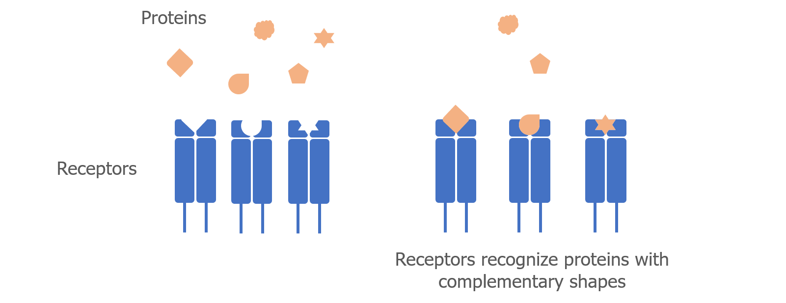
Immune cells obviously don’t have eyes or ears to see or hear trespassing pathogens. What they do have are proteins called receptors that interact with other proteins. The receptors have unique shapes that recognize proteins with complimentary shapes—similar to a lock that can only be opened by a specific key.

An immune response begins when an immune cell receptor (the “lock”) interacts with a protein from a pathogen (the “key”). A protein that is recognized by an immune cell receptor and triggers an immune response is called an antigen.

The receptors on innate immune cells recognize common proteins that are found on several types of pathogens, which gives them the flexibility to recognize more than one type of invader.

In contrast, the receptors on adaptive immune cells (T cells and B cells) recognize unique proteins that only certain types of pathogens have. This allows the immune system to “remember” a specific type of pathogen and to quickly begin an immune response if the pathogen infects the body again.

After the pathogen has been removed, the immune cells become deactivated and return to their normal state.

Many different diseases are caused by a malfunction of the normal immune response. This can happen in several ways. For example, immune cells may:

  • Fail to recognize a certain pathogen, which leads to chronic infection.
  • Mistakenly recognize a normal protein, which results in autoimmune disease.
  • Fail to “turn off” the immune response, which leads to chronic inflammation.

The next section will describe in more detail what goes wrong in autoimmune disease.

<< PREVIOUS                                                                                                                                         NEXT >>

GLOSSARY

Adaptive immune cells – Immune cells that recognize specific pathogens. Allow the immune system to “remember” pathogens in case of future infection.

Antigen – A protein that is recognized by an immune cell receptor and triggers an immune response.

Autoimmune disease – May occur when the immune system mistakenly recognizes a normal protein in the body as an invader and creates an immune response.

Immune cells – Specialized white blood cells (also called leukocytes) that fight infection.

Innate immune cells – Nonspecific immune cells. Recognize many pathogens. First responders to infection.

Pathogens – Bacteria and viruses that can cause disease.

Receptor – A protein that is located on the surface of a cell and interacts with other proteins. Receptors act as the “locks” that recognize specific pathogen “keys”.


RELATED

Share this Post

Immune Cells

Immune cells are specialized white blood cells (also called leukocytes) that circulate through our blood vessels and settle in many of our tissues and organs. They are on constant watch for anything that appears to be foreign and might be considered a threat to our health.

Immune cells are divided into two main groups—innate immune cells and adaptive immune cells. Within each of these groups are several subsets of cells that have specific jobs. Much like soldiers in a military operation, immune cells use different tactics and communication strategies to overpower the enemy.

Innate immune cells

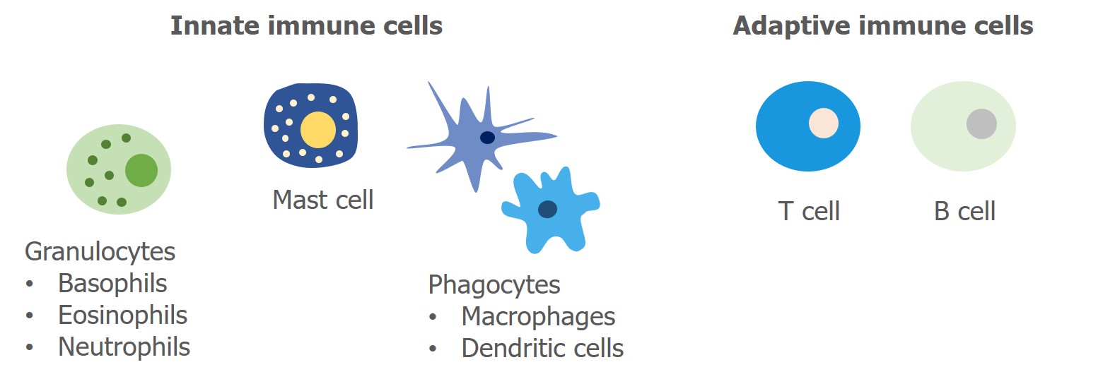
Innate immune cells act as the first responders to infection. They attack and eliminate many pathogens using basic methods that are harmful to cells. Different types of immune cells use different techniques. For example, cells called granulocytes (which include basophils, neutrophils, and eosinophils) kill pathogens by releasing toxic molecules. In contrast, other cells called phagocytes (which include macrophages and dendritic cells) ingest—or eat—pathogens to prevent them from causing damage to normal healthy tissues in the body.

Adaptive immune cells

Adaptive immune cells recognize specific pathogens and work together to create a customized immune response. Adaptive immune cells are either T lymphocytes (T cells) or B lymphocytes (B cells). These lymphocytes were named after the place in the body where they mature—T cells mature in the thymus and B cells in the bone marrow. T cells and B cells play major roles in autoimmunity and celiac disease and will be discussed in more detail in later sections.

<< PREVIOUS                                                                                                                                               NEXT >>

Glossary

Adaptive immune cells – Immune cells that recognize specific pathogens. Allow the immune system to “remember” pathogens in case of future infection.

B cell – A type of adaptive immune cell. Also called B lymphocyte.

Granulocytes – A type of innate immune cell that releases toxic molecules to fight infection.

Immune cells – Specialized white blood cells (also called leukocytes) that fight infection.

Innate immune cells – Nonspecific immune cells. Recognize many pathogens. First responders to infection.

Leukocytes – Specialized white blood cells (also called immune cells) that fight infection.

Lymphocytes – Adaptive immune cells. Can by B or T lymphocytes.

Pathogen – Bacteria and viruses that can cause disease.

Phagocytes – A type of innate immune cells that ingests (or eats) pathogens.

T cell – A type of adaptive immune cell. Also called T lymphocyte.


Related

Share this Post

The Immune System

Every day, potentially harmful bacteria and viruses (also called pathogens) try their best to infect us. Our immune system works tirelessly to defend against these attacks by preventing infection and killing pathogens before they cause harm.

The immune system is actually made up of many different parts, including:

  • Physical and chemical barriers (skin, lining of mouth and nose, mucus, enzymes)
  • Immune cells (white blood cells or leukocytes with specialized functions)
  • Cytokines (molecules that help immune cells communicate with each other)
  • Antibodies (molecules that recognize foreign pathogens)

When a pathogen gets past the physical and chemical barriers of the body, the immune cells are quickly called to action. Once activated, the immune cells produce molecules called cytokines and antibodies. By creating these molecules, immune cells are able to communicate with each other and destroy the invading pathogen.

The next section explains a bit more about the different types of immune cells in our bodies and the important jobs they have.

Immune Cells

Immune cells are specialized white blood cells (also called leukocytes) that circulate through our blood vessels and settle in many of our tissues and organs. They are on constant watch for anything that appears to be foreign and might be considered a threat to our health.

Immune cells are divided into two main groups—innate immune cells and adaptive immune cells. Within each of these groups are several subsets of cells that have specific jobs. Much like soldiers in a military operation, immune cells use different tactics and communication strategies to overpower the enemy.

Innate immune cells

Innate immune cells act as the first responders to infection. They attack and eliminate many pathogens using basic methods that are harmful to cells. Different types of immune cells use different techniques. For example, cells called granulocytes (which include basophils, neutrophils, and eosinophils) kill pathogens by releasing toxic molecules. In contrast, other cells called phagocytes (which include macrophages and dendritic cells) ingest—or eat—pathogens to prevent them from causing damage to normal healthy tissues in the body.

Adaptive immune cells

Adaptive immune cells recognize specific pathogens and work together to create a customized immune response. Adaptive immune cells are either T lymphocytes (T cells) or B lymphocytes (B cells). These lymphocytes were named after the place in the body where they mature—T cells mature in the thymus and B cells in the bone marrow. T cells and B cells play major roles in autoimmunity and celiac disease and will be discussed in more detail in later sections.

How Do Immune Cells Detect Pathogens?

Immune cells obviously don’t have eyes or ears to see or hear trespassing pathogens. What they do have are proteins called receptors that interact with other proteins. The receptors have unique shapes that recognize proteins with complimentary shapes—similar to a lock that can only be opened by a specific key.

An immune response begins when an immune cell receptor (the “lock”) interacts with a protein from a pathogen (the “key”). A protein that is recognized by an immune cell receptor and triggers an immune response is called an antigen.

The receptors on innate immune cells recognize common proteins that are found on several types of pathogens, which gives them the flexibility to recognize more than one type of invader.

In contrast, the receptors on adaptive immune cells (T cells and B cells) recognize unique proteins that only certain types of pathogens have. This allows the immune system to “remember” a specific type of pathogen and to quickly begin an immune response if the pathogen infects the body again.

After the pathogen has been removed, the immune cells become deactivated and return to their normal state.

Many different diseases are caused by a malfunction of the normal immune response. This can happen in several ways. For example, immune cells may:

  • Fail to recognize a certain pathogen, which leads to chronic infection.
  • Mistakenly recognize a normal protein, which results in autoimmune disease.
  • Fail to “turn off” the immune response, which leads to chronic inflammation.

The next section will describe in more detail what goes wrong in autoimmune disease.

NEXT TOPIC: What is autoimmune disease?

BACK TO MAIN PAGE

GLOSSARY

Adaptive immune cells – Immune cells that recognize specific pathogens. Allow the immune system to “remember” pathogens in case of future infection.

Antibodies – Y-shaped proteins that recognize foreign pathogens. Made by B cells. Also called immunoglobulins.

Antigen – A protein that is recognized by an immune cell receptor and triggers an immune response.

Autoimmune disease – May occur when the immune system mistakenly recognizes a normal protein in the body as an invader and creates an immune response.

B cell – A type of adaptive immune cell. Also called B lymphocyte.

Cytokines – Small proteins that are made and released by immune cells. Allows cells to send signals and provide instructions to other cells.

Granulocytes – A type of innate immune cell that releases toxic molecules to fight infection.

Immune cells – Specialized white blood cells (also called leukocytes) that fight infection.

Innate immune cells – Nonspecific immune cells. Recognize many pathogens. First responders to infection.

Leukocytes – Specialized white blood cells (also called immune cells) that fight infection.

Lymphocytes – Adaptive immune cells. Can by B or T lymphocytes.

Pathogens – Bacteria and viruses that can cause disease.

Phagocytes – A type of innate immune cells that ingests (or eats) pathogens.

Receptor – A protein that is located on the surface of a cell and interacts with other proteins. Receptors act as the “locks” that recognize specific pathogen “keys”.

T cell – A type of adaptive immune cell. Also called T lymphocyte.

Share this Post

GluTeen Free: Top “Fast Food” Restaurants

Here are some of our favorite “Fast Food” places to eat. We hope you enjoy them too.

Five Guys

Five Guys is one of the only fast food restaurants where you can get both a burger and fries without being too concerned about cross contamination, as the only food item that contains gluten in the entire facility are their gluten-filled buns. Their fries are 100% gluten free as they are the only items that enter their fryolators. Therefore, both the cajun fries and regular fries are gluten free! They do not have gluten free buns, but if you let the server know you have an issue with gluten, they will prepare your food in a separate area and will gladly either wrap your burger in lettuce or put it into a bowl. Milkshakes are definitely a no-go, however, as some can contain malted milk and/or Oreo mix-ins. Cross contamination in the blenders is highly possible. Also, avoid the fry sauce and (obviously) the malt vinegar on the tables.

Favorite Order: Bacon cheeseburger (wrapped in lettuce) with cajun fries.

Papa Gino’s

Even though many restaurants offer gluten-free pizza these days, very few are chains that can be found across the Northeast. When I found out that Papa Gino’s was serving pizzas on Udi’s gluten free crust, I was quite excited to know that I would be able to order a gluten free pizza for myself when getting pizza with my friends. They have a disclaimer on their website describing how they prepare these pizzas separately, use dedicated utensils/prep spaces and have trained their staff to know what toppings contain gluten (visit this link: https://www.papaginos.com/our-food/gluten-free). Additionally, pretty much all their toppings are gluten free aside from their meatballs, chicken tenders and mac-n-cheese (this is on pizzas?). TIP: Ask for the pizza well done, the crust is even better prepared this way.

Favorite Order: Plain cheese pizza or bacon pizza

Chipotle Mexican Grill

Whether you are traveling out of state or looking for a quick meal nearby, Chipotle is a convenient gluten-free-friendly Mexican fast food chain. There are many locations throughout the U.S. The only item containing gluten within the restaurant is the flour tortilla. So, as long as you request that your server changes their gloves and retrieves any cheese and lettuce you would like from out back (as gloves that may have handled the flour tortilla could have came in contact with these items) you should be all set! They offer gluten free corn tortillas, taco salads and burrito bowls as options.

Favorite Order: Burrito bowl with pork carnitas, cheese, lettuce, black beans, white rice, mild salsa and corn salsa

sweetgreen

This health conscious eatery has fourteen locations in Massachusetts, serving build-your-own salads and grain bowls. There is also a fixed menu (with gluten containing items marked with an asterisk) with different types of salads and bowls for those who are less creative. Everything in sweetgreen (at least according to their current menu- hopefully this remains to be the case!) is gluten free with the exception of their Za’atar Breadcrumbs which always seem to be located far from the rest of their assembly line to avoid the risk of cross contamination. Still ask your server to change their gloves just in case!

Favorite Order: Toss up between the Kale Caesar or Pesto Portabello

The GluTeen Free blog posts are written by Sarah and Kaitlyn Roffman, twins with celiac disease who are currently juniors at Littleton High School.